CLEAR item#44
“Baseline demographic and clinical characteristics. Provide the baseline demographic, clinical, and imaging characteristics in text and/or tables. Report the information separately for training, validation (i.e., cross- validation), and test datasets, along with grouping based on the reference standard or non-radiomic variables. Associated statistical tests should also be provided to identify if the sets are identical or not. Provide whether any confounder is detected and handled appropriately.” [1] (from the article by Kocak et al.; licensed under CC BY 4.0)
Reporting examples for CLEAR item#44
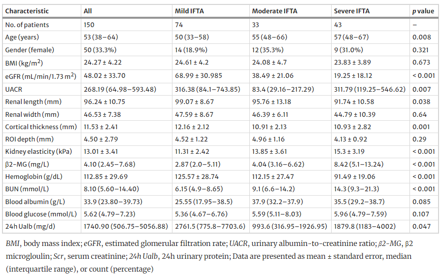
Example#1. “A total of 150 patients with CKD were identified in Table 1” [2] (from the article by Ge et al.; licensed under CC BY 4.0)
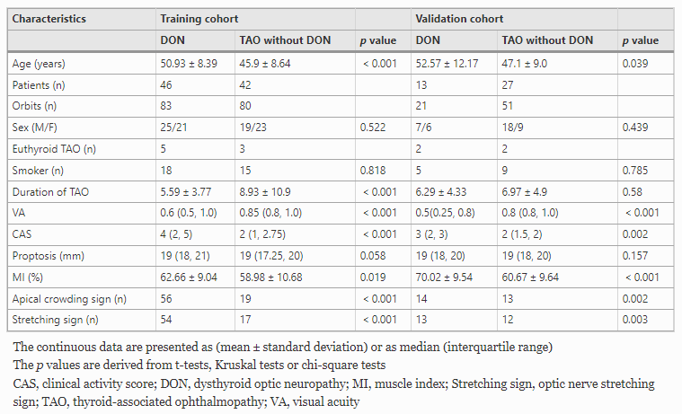
Example#2. “Table 1 shows the detailed demographic information of the participants” [3] (from the article by Wu et al.; licensed under CC BY 4.0)
Explanation and elaboration of CLEAR item#44
In Example#1, a table is used to present demographic, ultrasound, and laboratory findings for 150 patients with chronic kidney disease (CKD). This presentation aids in understanding the population’s characteristics and the context of the study. Example#2 shows demographic information, distinguishing between training and test sets. This separation is crucial for assessing the balance of the datasets, ensuring that the training and test data are comparable.
References
- Kocak B, Baessler B, Bakas S, et al (2023) CheckList for EvaluAtion of Radiomics research (CLEAR): a step-by-step reporting guideline for authors and reviewers endorsed by ESR and EuSoMII. Insights Imaging 14:75. https://doi.org/10.1186/s13244-023-01415-8
- Ge X-Y, Lan Z-K, Lan Q-Q, et al (2023) Diagnostic accuracy of ultrasound-based multimodal radiomics modeling for fibrosis detection in chronic kidney disease. Eur Radiol 33:2386–2398. https://doi.org/10.1007/s00330-022-09268-3
- Wu H, Luo B, Zhao Y, et al (2022) Radiomics analysis of the optic nerve for detecting dysthyroid optic neuropathy, based on water-fat imaging. Insights Imaging 13:154. https://doi.org/10.1186/s13244-022-01292-7